Nuffield Theatre
University of Southampton
09 July - 11 July, 2018
Southampton, United Kingdom

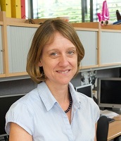
Professor Alison Noble
Institute of Biomedical Engineering
University of Oxford, U.K.
Biography
Professor Alison Noble is the Technikos Professor of Biomedical Engineering in the Oxford University
Department of Engineering Science, Associate Head of MPLS Division (Industry and Innovation), and a Fellow of St Hilda's College,
Oxford. She is a former Director of the Institute of Biomedical Engineering (2012-16). Professor Noble is a Fellow of the Royal Society,
Fellow of the IET, a Fellow of the MICCAI Society, and a Fellow of the Royal Academy of Engineering. She is the current Chair of the
EPSRC Healthcare Technologies Strategic Advisory Team.
Title: How Machine Learning Is Changing Ultrasound Image Analysis
Abstract: We hear a lot about machine learning applied to medical imaging. Much of the current focus is on
automated image segmentation and object recognition. But is it a matter of taking software architectures published
in computer vision and re-training with medical images as input, or are there specific challenges and solutions
which are specific to the medical imaging domain which require a different approach? In this talk, I will use
on-going research in my laboratory to illustrate how we are making modality-specific advances in machine learning
applied to ultrasound imaging. Examples will be drawn from western world and LMIC (low and middle income country)
applications of medical ultrasound.

Professor Tom Wilkinson
NIHR Southampton Biomedical Research Centre
University Hospital Southampton NHS Foundation Trust, U.K.
Biography
Professor Tom Wilkinson is a Professor of Respiratory Medicine at the University of Southampton, Faculty of Medicine and Honorary
Consultant at University Hospitals Southampton. As a clinician and researcher interested in asthma and chronic obstructive pulmonary disease (COPD),
Professor Wilkinson leads the research into airway diseases. He is also lead of the Southampton COPD research group,
and respiratory theme lead for the NIHR Wessex CLAHRC. Close collaborations in Southampton, London and Oxford have been
established to uncover the interaction between nutritional status, immune responses and infection. He has contributed
to the establishment of unique ex-vivo and in vivo infection challenge models and is clinical lead on the EVITA programme.

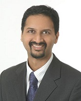
Professor Anant Madabhushi
Centre of Computational Imaging and Personalized Diagnostics
Case Western Reserve University, U.S.
Biography
Dr. Anant Madabhushi is the F. Alex Nason professor II of biomedical engineering at Case Western Reserve University in Cleveland and director
of the university’s Center for Computational Imaging and Personalized Diagnostics (CCIPD).He leads to develop tools to computationally interrogate
biomedical image data, searching for patterns and connections that will help in disease diagnosis, prognosis and theragnosis.
Dr. Madabhushi currently holds 24 patents in medical imaging analysis, computer vision and computer-aided diagnosis. In 2017, he was
awarded patents for “Histogram of hosoya index (HoH) features for quantitative histomorphometry” and “Textural analysis of lung nodules.”
Dr. Madabhushi is a Wallace H. Coulter Fellow and a fellow of the American Institute of Medical and Biological Engineering (AIMBE).
Title: Computational Imaging and Artificial Intelligence: Implications for Precision Medicine
Abstract: Traditional biology generally looks at only a few aspects of an organism at a time and attempts to molecularly dissect diseases and
study them part by part with the hope that the sum of knowledge of parts would help explain the operation of the whole. Rarely has this been a
successful strategy to understand the causes and cures for complex diseases. The motivation for a systems based approach to disease understanding
aims to understand how large numbers of interrelated health variables, gene expression profiling, its cellular architecture and microenvironment,
as seen in its histological image features, its 3 dimensional tissue architecture and vascularization, as seen in dynamic contrast enhanced (DCE)
MRI, and its metabolic features, as seen by Magnetic Resonance Spectroscopy (MRS) or Positron Emission Tomography (PET), result in emergence of
definable phenotypes. At the Center for Computational Imaging and Personalized Diagnostics (CCIPD) at Case Western Reserve University, we have been
developing computerized knowledge alignment, representation, and fusion tools for integrating and correlating heterogeneous biological data spanning
different spatial and temporal scales, modalities, and functionalities. These tools include computerized feature analysis methods for extracting
subvisual attributes for characterizing disease appearance and behavior on radiographic (radiomics) and digitized pathology images (pathomics).
In this talk I will discuss the development work in CCIPD on new radiomic and pathomic approaches for capturing intra-tumoral heterogeneity and
modeling tumor appearance. I will also focus my talk on how these radiomic and pathomic approaches can be applied to predicting disease outcome,
recurrence, progression and response to therapy in the context of prostate, brain, rectal, oropharyngeal, and lung cancers. Additionally I will
also discuss some recent work on looking at use of pathomics in the context of racial health disparity and creation of more precise and tailored
prognostic and response prediction models.