• Nuffield Theatre
    University of Southampton


    09 July - 11 July, 2018
    Southampton, United Kingdom

  • programme

    Monday 09th July 2018
    09.00-10.00 Registration
    10.00-11.00 Deep Learning in Medical Imaging
    Chair: Ingela Nystrom
    - N. Alsubaie, M. Shaban, D. Snead, A. Khurram, N. Rajpoot, A Multi-Resolution Deep Learning Framework for Lung Adenocarcinoma Growth Pattern Classification.
    - S. Agarwala, A. Kumar, D. Nandi, A. Dhara, A. Sadhu, S. Thakur, A. Bhadra, Convolutional Neural Networks for Efficient Localization of Interstitial Lung Disease Patterns in HRCT Images.
    - Y. Almutairi, T. Cootes, K. Kadler, Segmentation Nucleus Membranes in SBFSEM Volume Data with Deep Neural Networks.
    11.00-11.30 Coffee break
    11.30-12.30 Keynote 1: Prof. Alison Noble, University of Oxford
    How Machine Learning Is Changing Ultrasound Image Analysis
    Chair: Mark Nixon
    12.30-13.30 Lunch break
    13.30-14.50 Special session: Liver Analysis
    Chair: Constantino Reyes Aldasoro
    - K. Arndtz, B. Irving, P. Eddowes, D. Green, M. Kelly, N. Jayaratne, R. Banerjee, J. M.l Brady, G. M. Hirschfield, Novel quantitative magnetic resonance imaging features with liver function tests to distinguish parenchymal and biliary disease.
    - B. Irving, C. Hutton, K. Arndtz, N. Jayaratne, M. Kelly, R. Banerjee, G. M. Hirschfield, J. M. Brady, Regional assessment of liver disease progression and response to therapy by multi-time point m-SLIC correspondence.
    - M. Wojciechowska, B. Irving, A. Dennis, H. Wilman, R. Banerjee, M. Brady, M. Kelly, Automated detection of cystic lesions in quantitative T1 liver images.
    - G. Ridgway, K. Janowski, A. Dennis, V. Bachtiar, J. McGonigle, C. Everitt, A. Darekar, D. Breen, D. Green, S. Neubauer, R. Banerjee, P. Socha, M. Brady, Voxel-wise analysis of paediatric liver MRI.
    14.50-15.05 Poster highlights
    Chair: Mark Nixon
    - E. Abouei, A. Lee, G. Hohert, P. Lane, S. Lam, C. MacAulay, Quantitative Evaluation of Correction Methods and Simulation of Motion Artifacts for Rotary Pullback Imaging Catheters.
    - Z. Suhail, A. Mahmood, L. Wang, P. Malcolm, R. Zwiggelaar, A Voting-Based Encoding Technique for the Classification of Gleason Score for Prostate Cancers.
    - A. Kumar, S. Agarwala, A. Dhara, D. Nandi, S. Thakur, A. Sadhu, A. Bhadra, Segmentation of Lung Field in HRCT Images using U-Net Based Fully Convolutional Networks.
    - V. Azzopardi, M. Guy, E. Lewis, Identifying shape-based biomarkers for diagnosis of Parkinson’s Disease from Ioflupane (123I) SPECT data.
    - M. Fan, J. Vicory, S. McGill, S. Pizer, J. Rosenman, Features for the Detection of Flat Polyps in Colonoscopy Video.
    - R. Mukundan, Feature Analysis of Biomarker Descriptors for HER2 Classification of Histology Slides.
    - J. Verhoeven, N. Miller, C. C. Reyes-Aldasoro, Analysis of the symmetry of electrodes for Electropalatography with Cone Bean CT Scanning.
    15.05-15.40 Tea break (and posters)
    15.40-16.40 Texture and Image Analysis (Session 1)
    Chair: Mike Brady
    - M. George, R. Zwiggelaar, Breast Tissue Classification using Local Binary Pattern variants: A Comparative study.
    - Y. Almakady, S. Mahmoodi, J. Conway, M. Bennett, Volumetric Texture Analysis based on Three-Dimensional Gaussian Markov Random Fields for COPD Detection.
    - O. Leonovych, M. R. Koujan, A. Akram, J. Westerfeld, D. Wilson, K. Dhaliwal, S. McLaughlin, A. Perperidis, Texture descriptors for classifying sparse, irregularly sampled optical endomicroscopy images.
    16.40-16.50 Sponsor Presentations
    Chair: Sasan Mahmoodi
    - MathWorks
    - MedIAN
    16.50-17.30 Texture and Image Analysis (Session 2)
    Chair: Mike Brady
    - D. Farnell, J. Galloway, A. Zhurov, S. Richmond, P. Pirttiniemi, R. Lähdesmäki, What’s in a Smile? Initial Results of Multilevel Principal Components Analysis of Facial Shape and Image Texture.
    - A. Alfahaid, T. Morris, An automated age-related macular degeneration classification based on local texture features in optical coherence tomography angiography.
    19.00 Welcome reception
    Tuesday 10th July 2018
    09.00-10.00 MRI: Applications and Techniques
    Chair: Maria Valdes Hernandez
    - F. Lennartsson, A. Darekar, K. Maharatna, D. Konn, D. Allen, J. Tournier, J. Broulidakis, B. Vollmer, Developing a framework for studying brain networks in neonatal hypoxic-ischemic encephalopathy.
    - M. Brudfors, Y. Balbastre, P. Nachev, J. Ashburner, MRI Super-Resolution using Multi-Channel Total Variation.
    - P. Stroe, X. Xie, A. Paiement, Manifold modeling of the beating heart motion.
    10.00-10.30 Coffee break
    10.30-11.30 Keynote 2: Prof. Anant Madabhushi, Case Western Reserve University
    Computational Imaging and Artificial Intelligence: Implications for Precision Medicine
    Chair: Nasir Rajpoot
    11.30-12.30 Segmentation in Medical Images
    Chair: Yalin Zheng
    - C. Karabag, M. Jones, C. Peddie, A. Weston, L. Collinson, C. C. Reyes-Aldasoro, Automated Segmentation of HeLa Nuclear Envelope from Electron Microscopy Images.
    - N. Alam, A. Oliver, E. Denton, R. Zwiggelaar, Automatic Segmentation of Microcalcification Clusters.
    - J. A. Solis-Lemus, B. Stramer, G. Slabaugh, C. C. Reyes-Aldasoro, Analysis of the Interactions of Migrating Macrophages.
    12.30-13.30 Lunch break
    13.30-14.30 Matlab
    Chair: Mark Nixon
    14.30-14.50 Clinical abstracts
    Chair: Emma Lewis
    - L. Vass, M. Fisk, M. Polkey and I. Wilkinson, Compartmental modelling of 18F-FDG to assess Pulmonary Inflammation in Obstructive Sleep Apneoa/Hypopneoa Syndrome (OSAHs) Patients.
    - L. Ballerini, M. C. V. Hernández, R. Lovreglio, S. M. Maniega, M. E. Bastin, I. J. Deary, J. M. Wardlaw, Clinical Validation of Automatic Quantitative Measurements of Perivascular Spaces in Older Age MRI Brain Data.
    - J. A. Leighs, E. B. Lewis, A. A. Darekar, E. Scorletti, C. D. Byrne, Searching for Non-Alcoholic Fatty Liver Disease (NAFLD) Biomarkers in Magnetic Resonance Imaging.
    - M. C. V. Hernández, L. Ballerini, A. Gow, A. Glatz, S. M. Maniega, M. E. Bastin, I. J. Deary, J. Starr, J. M. Wardlaw, Perivascular spaces in the centrum semiovale automatically assessed in MRI, their association with total brain iron deposition and effect on cognition in the elderly.
    14.50-15.15 Tea break (and clinical abstracts)
    15.15-16.15 CT: Learning and Planning
    Chair: Tryphon Lambrou
    - L. Berger, E. Hyde, M. Jorge Cardoso, S. Ourselin, An Adaptive Sampling Scheme to Efficiently Train Fully Convolutional Networks for Semantic Segmentation.
    - M. Daykin, M. Sellathurai, I. Poole, A Comparison of Unsupervised Abnormality Detection Methods for Interstitial Lung Disease.
    - L. Blache, F. Nysjö, F. Malmberg, A. Thor, A. Lorenzo, I. Nystrom, SoftCut: a virtual planning tool for soft tissue resection on CT images.
    16.15-17.15 Journal Paper Writing Club
    19.00 Drinks reception
    20.00 Conference dinner
    Wednesday 11th July 2018
    09.00-10.00 Special session: Ocular Imaging Analysis
    Chair: Xujiong Ye
    - M. Badar, M. Shahzad, M. M. Fraz, Simultaneous Segmentation of Multiple Retinal Pathologies Using Fully Convolutional Deep Neural Network.
    - Y. Xu, B. Williams, B. Al-Bander, Z. Yan, Y. Shen, Y. Zheng, Improving the Resolution of Retinal OCT with Deep Learning.
    - X. Xu, R. Wang, T. Tan, F. Xu, An improved U-Net architecture for simultaneous arteriole and venule segmentation in fundus image.
    10.00-11.00 Keynote 3: Prof. Tom Wilkinson, University of Southampton
    Chair: Mark Nixon
    11.00-11.30 Coffee break
    11.30-12.30 Applications of Medical Image Analysis
    Chair: Yalin Zheng
    - E. Kugler, T. Chico, P. Armitage, Image Analysis in Light Sheet Fluorescence Microscopy Images of Transgenic Zebrafish Vascular Development.
    - A. N. Fatihah, F. Delbressine, L. Feijs, S. B. Oetomo, Color Correction of Baby Images for Cyanosis Detection.
    - R. Awan, N. Rajpoot, Deep Autoencoder Features for Registration of Histology Images.
    12.30-13.30 Lunch break

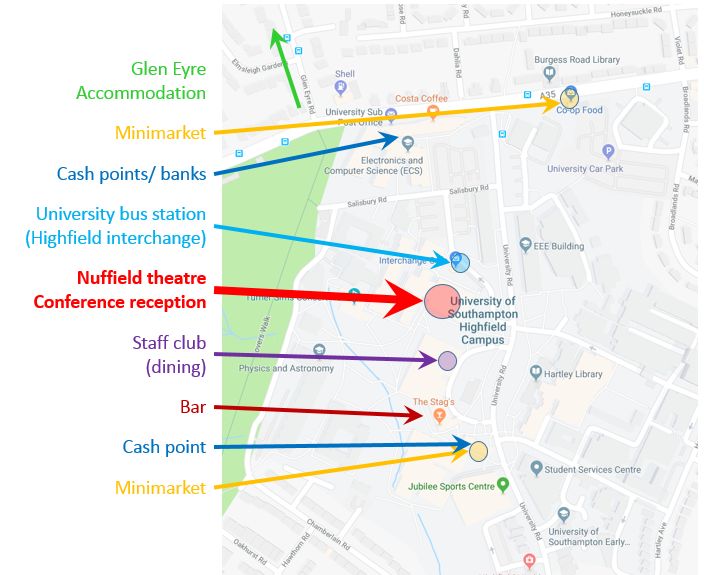
    Please refer to the site map of the Highfield Campus for useful information.